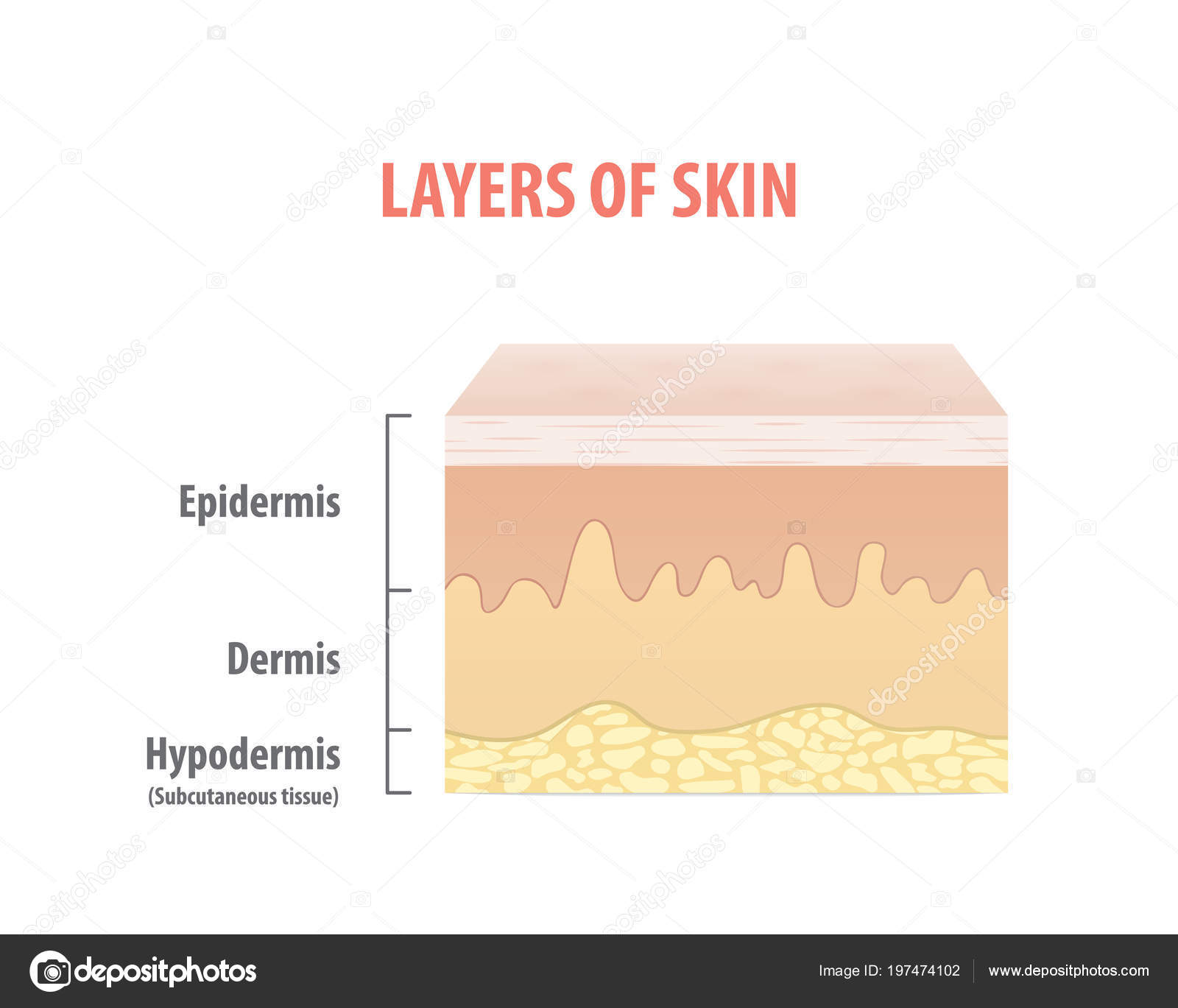
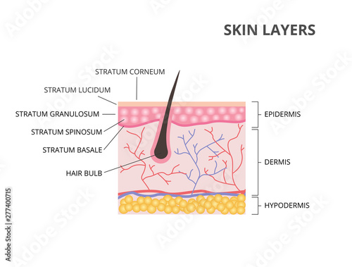
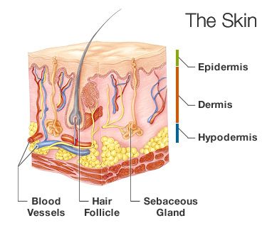
Skin Layers Diagram
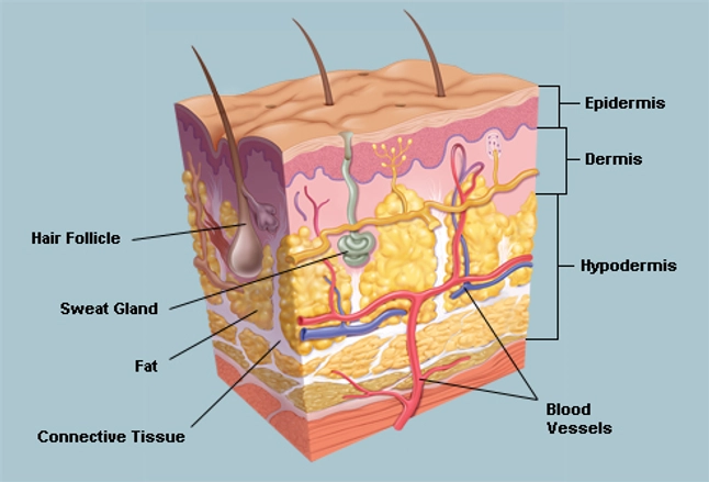
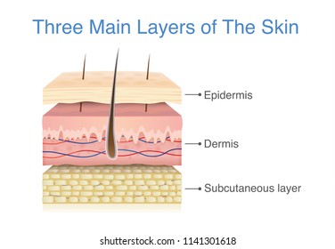
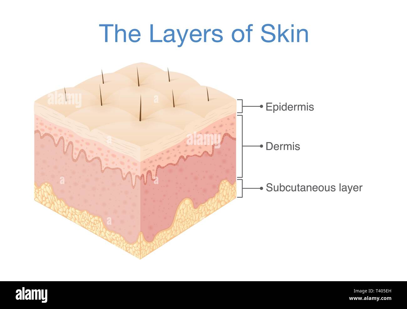
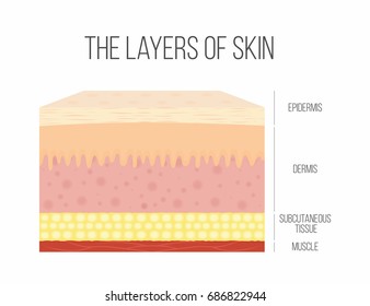
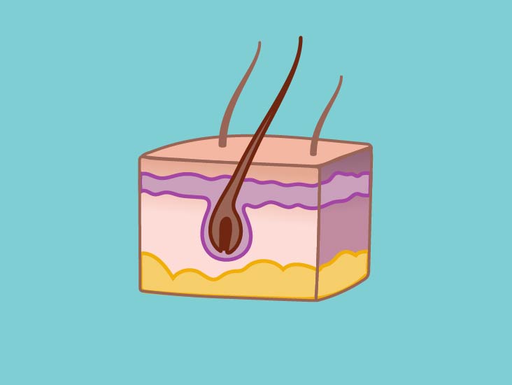
The epidermis the dermis and subcutaneous tissue. The skin is composed of two main layers.
 Layers Of Skin How Many Diagram Model Anatomy In Order
The human skin is the outer covering of the body and is the largest organ of the integumentary system.

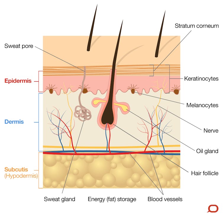
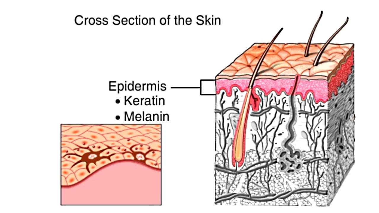
Skin layers diagram. Human skin in human anatomy the covering or integument of the bodys surface that both provides protection and receives sensory stimuli from the external environment. Skin has two main layers both of which serve a purpose. The cells on the surface are constantly falling off shedding.
Human skin is similar to most of the other mammals skin and it is very similar to pig skin. The epidermis is the outermost layer of the three layers of skin. Other animal coverings such as the arthropod exoskeleton have different developmental origin structure and chemical composition.
We will now go over the skins layers in more detail. This is called. As can be seen in the skin diagram the outermost layer of the skin is called the epidermis layer.
The skin is composed of two main layers. Webmds skin anatomy page provides a detailed image of the skin and its parts as well as a medical definition. Beneath the two layers is a layer of subcutaneous fat which also protects your body and helps you adjust to.
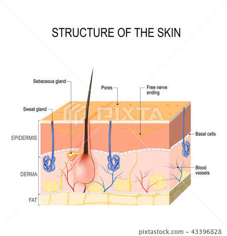
The epidermis is the layer of skin that we can see. The epidermis an outermost layer that contains the primary protective structure. Structure of the skin.
Skin is the soft outer tissue covering of vertebrates with three main functions. The skin consists of three layers of tissue. There are no blood vessels in the epidermis but its deepest layer is supplied with lymph fluid.
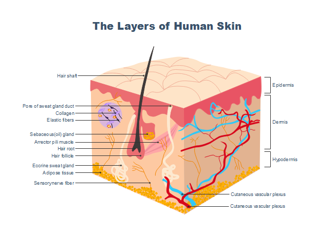
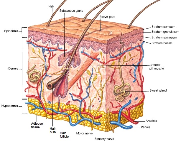
The adjective cutaneous means of the skin from latin cutis skin. The skin has up to seven layers of ectodermal tissue and guards the underlying muscles bones ligaments and internal organs. This skin diagram clearly shows all the layers of skin.
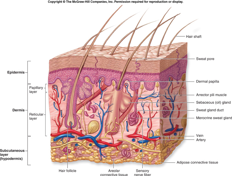
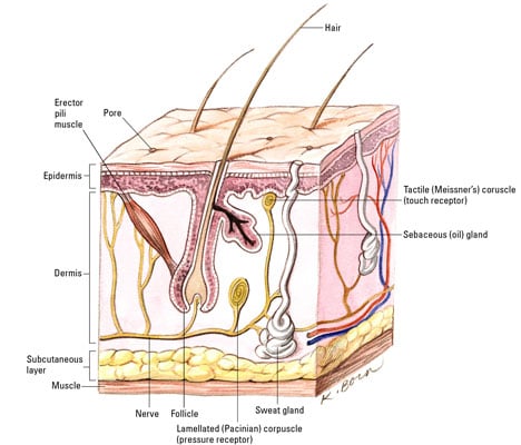
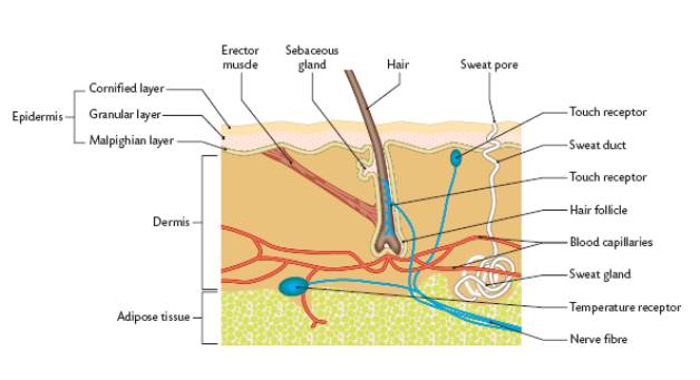
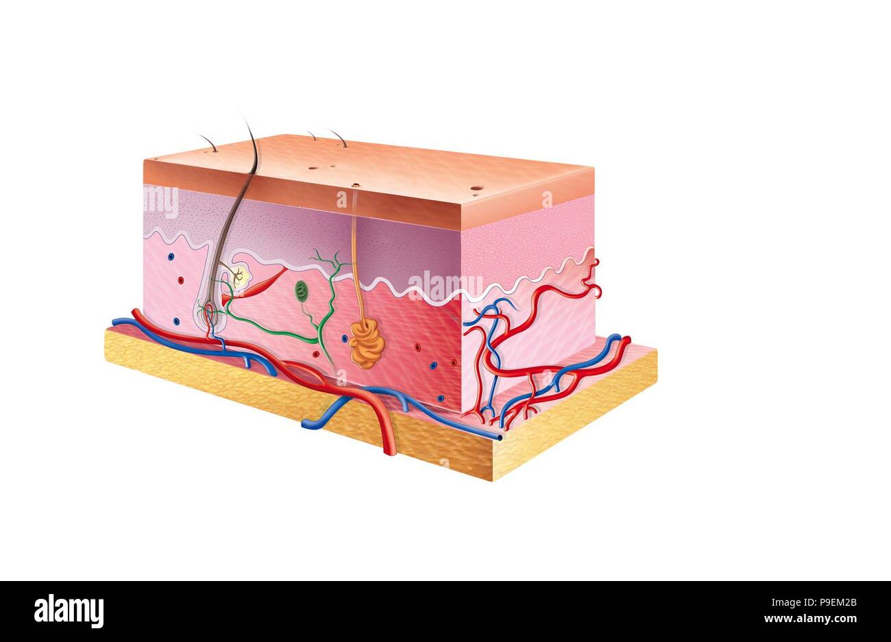
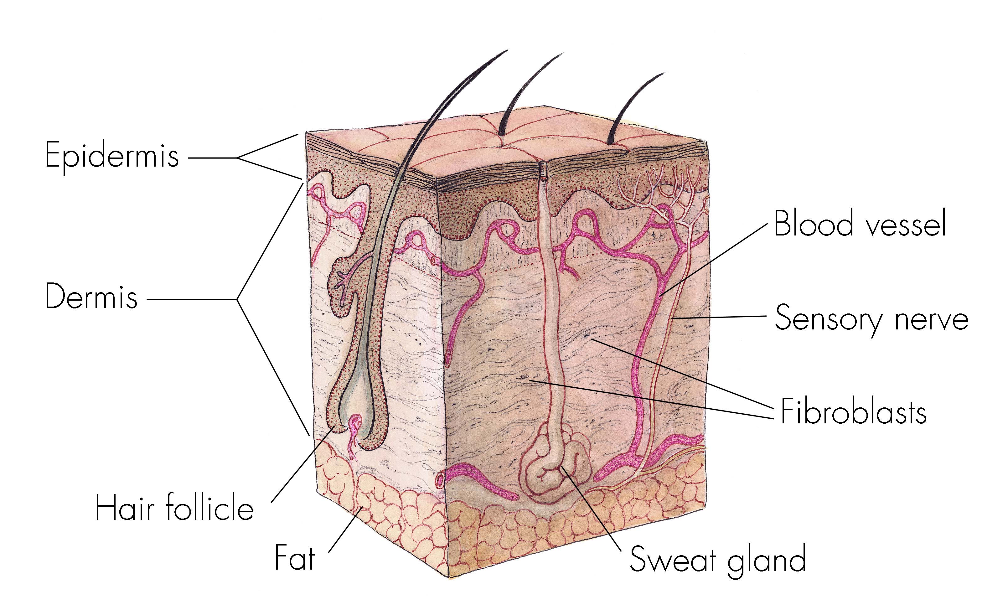
The epidermis made of closely packed epithelial cells and the dermis made of dense irregular connective tissue that houses blood vessels hair follicles sweat glands and other structures. Verywell alexandra gordon the epidermis. The three layers of skin.
Its thickness depends on where it is located on the body. It varies in thickness depending on the part of the body it is thickest on the soles of the feet and palms of the hand and thinnest on eyelids and nipples. Learning how the skin functions begins with an understanding of the structure of the three layers of skin.
Protection regulation and sensation. The epidermis made of closely packed epithelial cells and the dermis made of dense irregular connective tissue that houses blood vessels hair follicles sweat glands and other structures. Learn about the skins function and conditions that may affect the skin.
What is the epidermis.
 The Skin Human Anatomy Picture Definition Function And
 Diagram Of The Human Skin Layers Skin Structure
 5 1 Layers Of The Skin Anatomy And Physiology
 Understand How The Skin Layers Work For Repair
 Skin Layers Diagram Best Natural Skin Care Skin
 The Skin Series Part I Functions Anatomy Skincare Academy
 Sketch Of The Human Skin Layers Moving From The Outside To
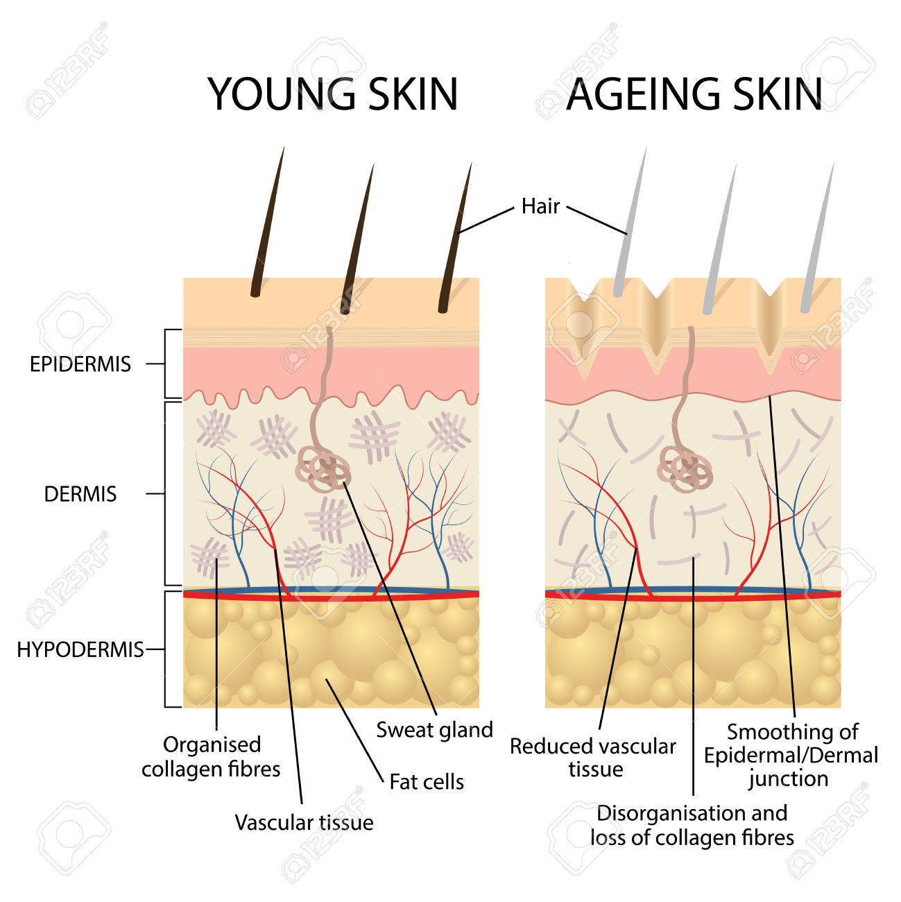 Young Healthy Skin And Older Skin Comparison Skin Layers And
 Skin Structure And Function Explained
 1 Skin Layers Source 17 Download Scientific Diagram
 Berkas Skin Layers Svg Wikipedia Bahasa Indonesia
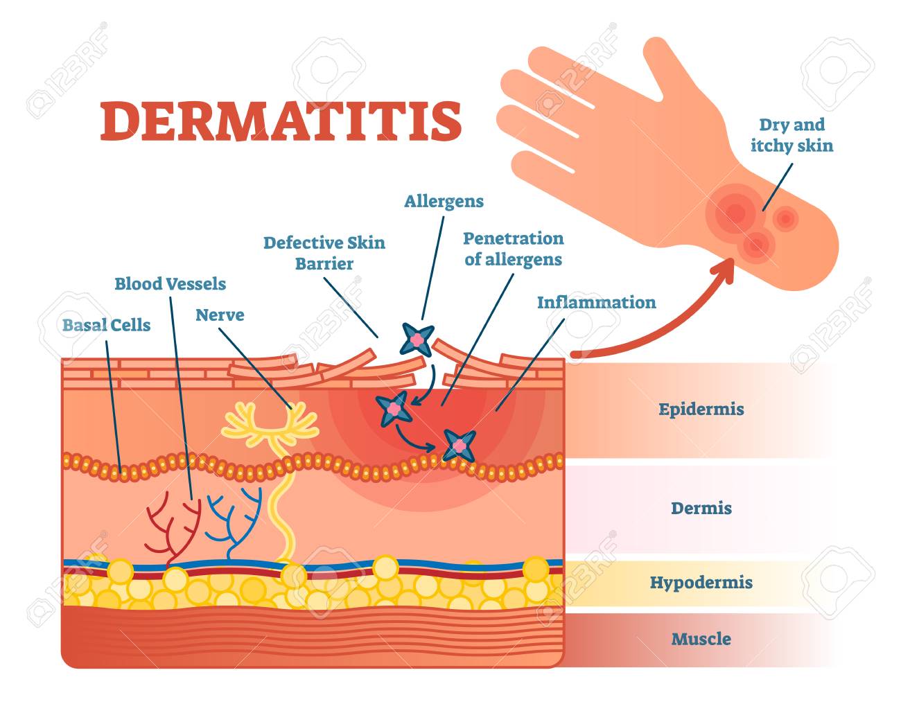 Dermatitis Flat Vector Illustration Diagram With Skin Layers
 Layers Of The Epidermis Layers Of The Epidermis Layers Of
 Vectores Imagenes Y Arte Vectorial De Stock Sobre Human
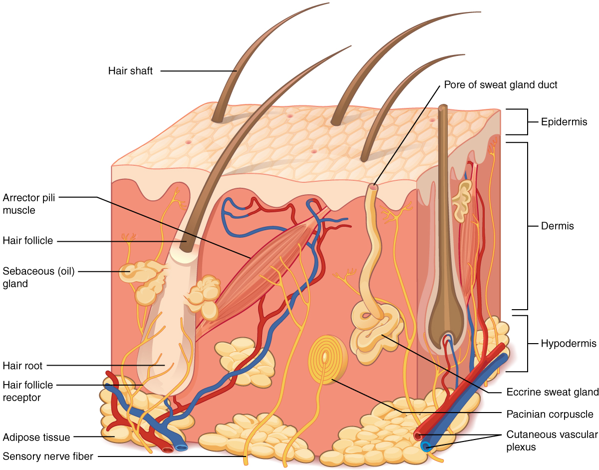
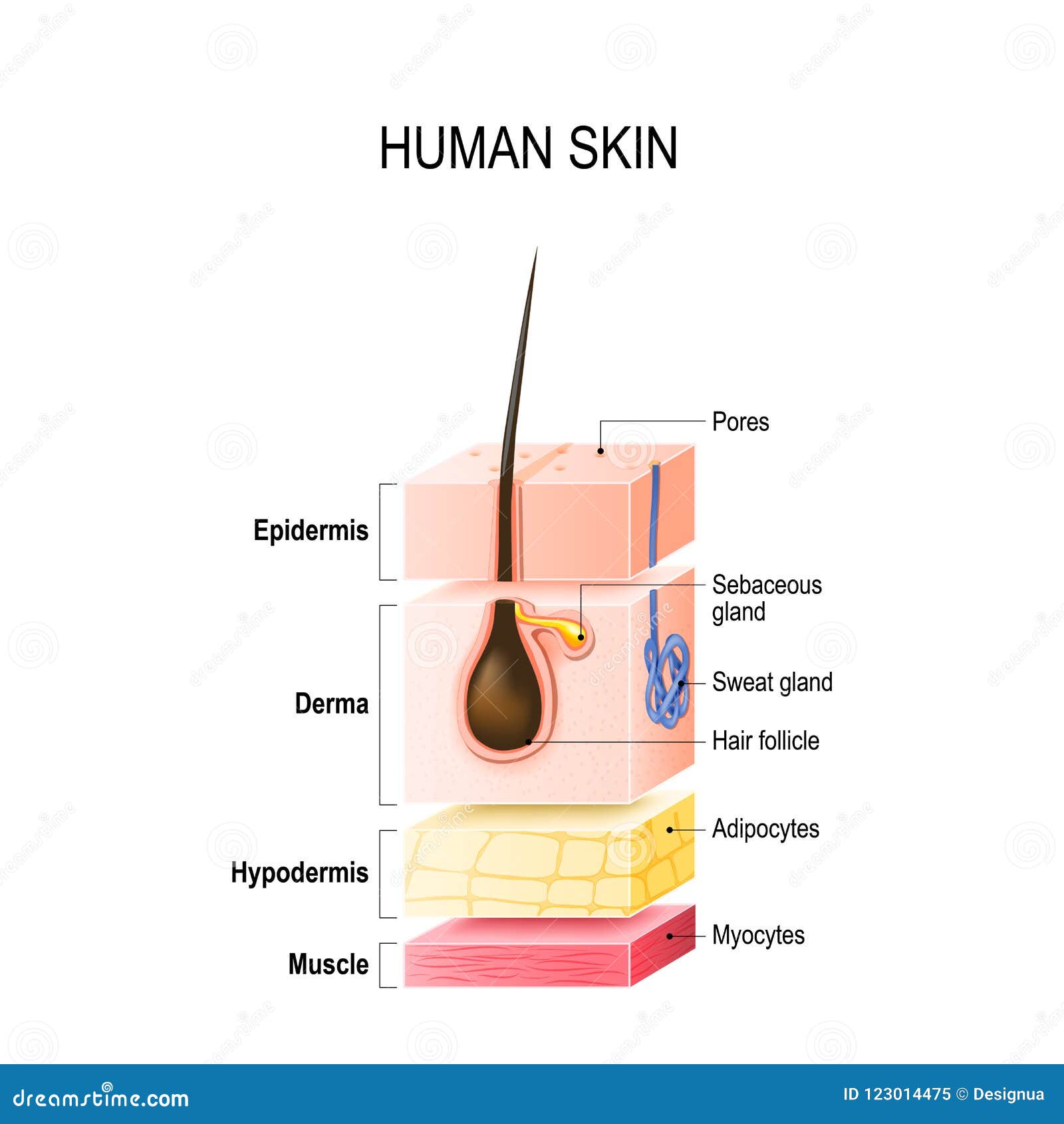
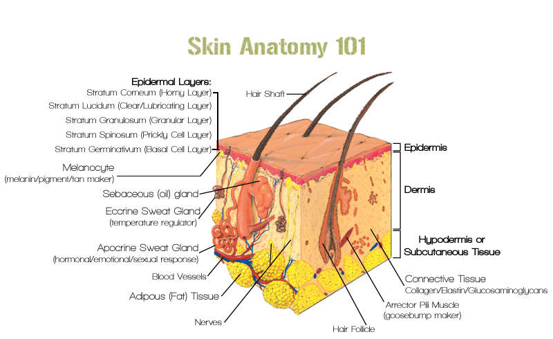
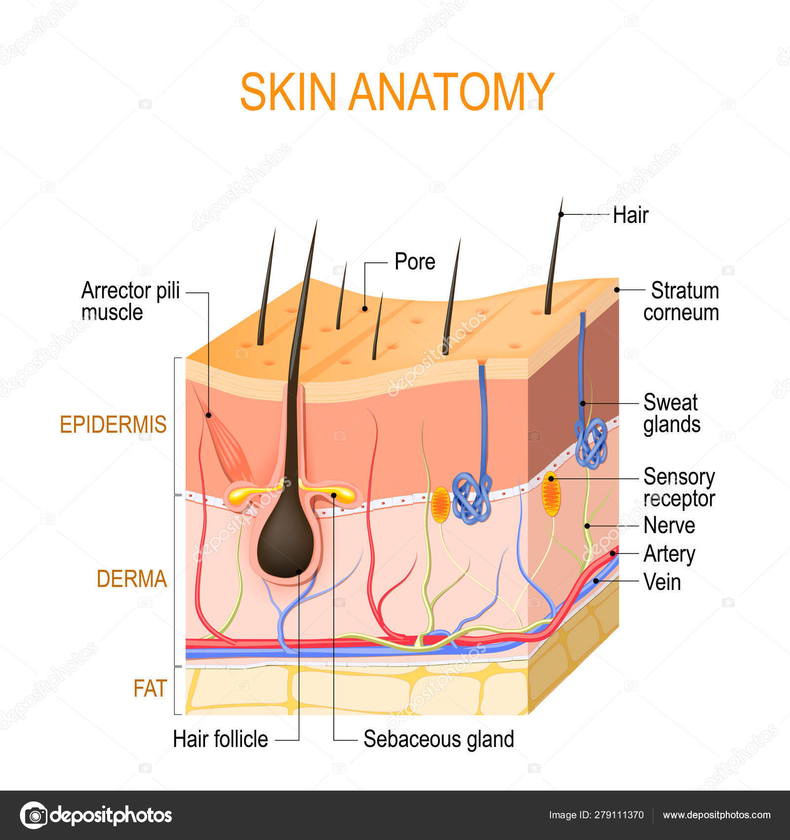 Skin Anatomy Layers Epidermis With Hair Follicle Sweat
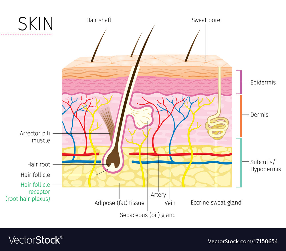 Human Anatomy Skin And Hair Diagram
 Layers Of Skin Skincare Face Treatments Ent Wellbeing Sydney
 The Anatomy Of Skin Dummies
 Skin Layers Diagram Quizlet
 Imagenes Fotos De Stock Y Vectores Sobre Layers Dermis
 Free Human Skin Layers Templates
:max_bytes(150000):strip_icc()/skin-anatomy-1068880_review-01-9adf9daebac8464eb693274a960bd850.png) Skin Anatomy The Layers Of Skin And Their Functions
Skin Anatomy The Layers Of Skin And Their Functions
 Schematic Representation Of Skin Layer Download
 Skin Layers Epidermis Dermis Hypodermis Isometric Vector Illustration
 Layers Of Normal Human Skin Stock Vector Illustration Of
 Gen A P Lab 10 The Skin Layers Of The Skin Diagram Quizlet
 Skin Layers Vector Photo Free Trial Bigstock

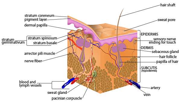 Seer Training Anatomy Of The Skin
 Skin Layers Diagram Google Search All Things Skin Skin
 Human Skin Anatomy Image Photo Free Trial Bigstock
 Layers Skin Diagram Illustration Vector White Background
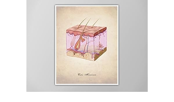 Amazon Com Human Skin Human Anatomy Art Print Human
![]() Layers Of Human Skin Melanocyte And Melanin Stock Vector
Layers Of Human Skin Melanocyte And Melanin Stock Vector
 Does Anyone Know How Thick The Skin Epidermis And Dermis
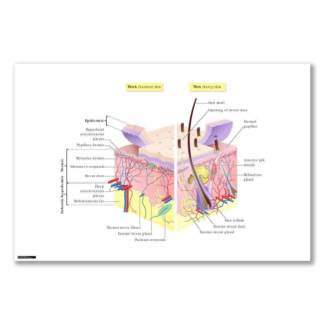 Amazon Com Ldgsfdsdsa Skin Layers Diagram Thick Thin
 A Diagram Layers Human Skin Two Main Layers Are Epidermis

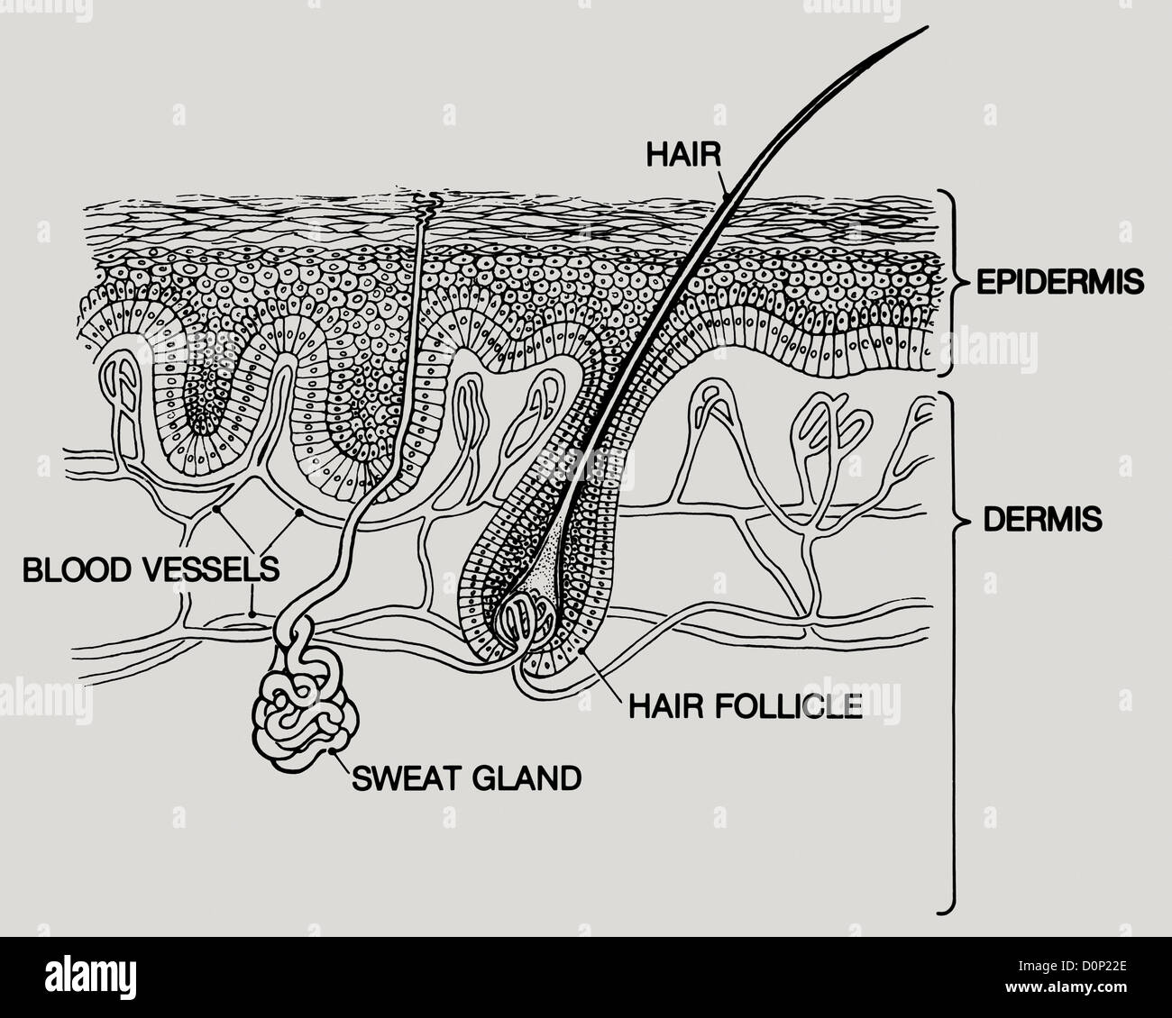
 Diagram Of A Hair Follicle In A Cross Section Of Skin Layers
 Young And Older Skin
 Human Skin Atlas
 Young Healthy Skin And Older Skin Comparison
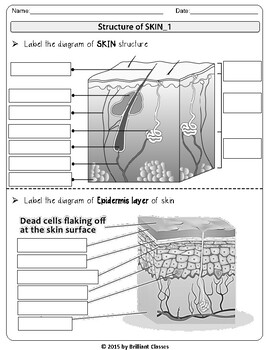
 28 Skin Diagram Anatomy Blank Skin Structure Diagram
![]() Diagram Of A Hair Follicle In A Cross Section Of Skin Layers
Diagram Of A Hair Follicle In A Cross Section Of Skin Layers
 Show Pictures Integumentary System This Skin Diagram
 Different Skin Layers Download Scientific Diagram


 Epidermis Layers Epithelial Cells Structure Of The Humans Skin
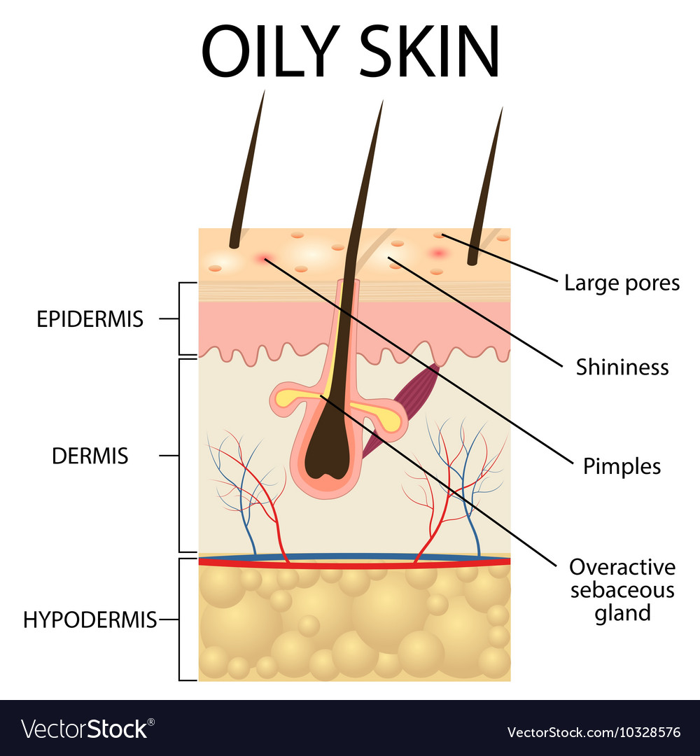 The Layers Of Oily Skin
 28 7 Layers Of Skin Diagram Layers Of Skin Related
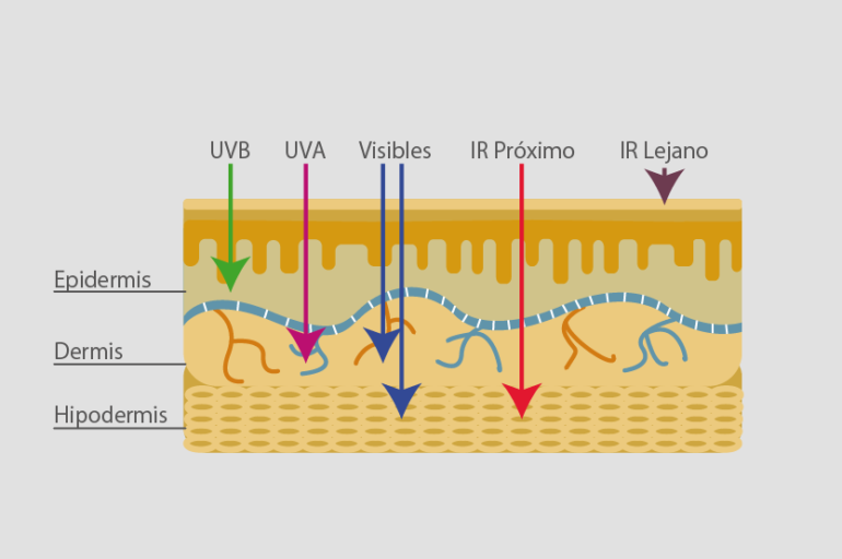 How The Sun Affects The Different Skin Layers Vitae Health
 Skin Layers Epidermis Dermis Hypodermis Flat Vector
 Human Skin Diagram Stock Photos Human Skin Diagram Stock
 Diagram Of Human Skin Layers Charlotte Desire
 Human Skin Anatomy Art Print Skin Layers Skin Diagram Art
 Diagram Of A Hair Follicle In A Cross Section Of Skin Layers

 The Skin Boundless Anatomy And Physiology
 The Skin Is A Very Important And Our Largest Organ What

![]() Back Skin Diagram Wiring Diagram Rows
Back Skin Diagram Wiring Diagram Rows
 Perspiring Anatomical Skin Cross Section Vector Illustration
 Structure Of The Skin In Dogs Dog Owners Veterinary Manual
 Skin Layers Healthy Normal Human Skin
 Skin Diagram Layers Diagram
 Skin Layers Stock Photo 212379395 Alamy
 Printable Skin Diagram To Label Owner Manual Wiring Diagram
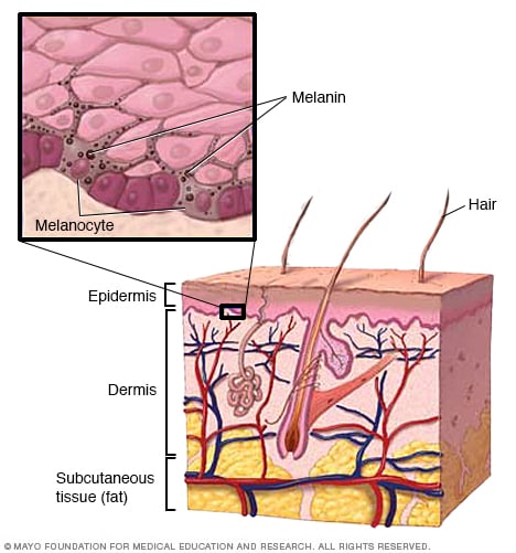 Skin Layers And Melanin Mayo Clinic
 Skin Layers With Sebaceous Gland And Sweat Glands Stock
 Skin Diagram Worksheet Have Fun Teaching
 Mechanistic Multilayer Model For Non Invasive Bioimpedance
Structure Of The Skin Course Hero
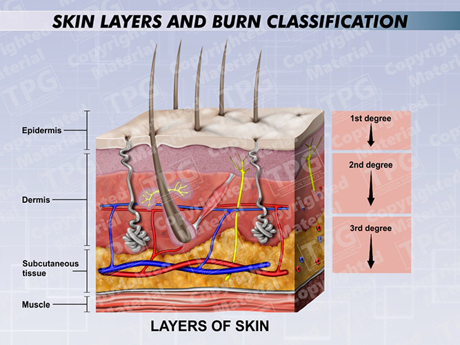 Skin Layers And Burn Classification Order
 Young Healthy Sking Vector Photo Free Trial Bigstock
 Young And Older Skin Stock Illustration I5427316 At Featurepics
 Imagenes Fotos De Stock Y Vectores Sobre Skin Diagram
 Schematic Diagram Of Skin Layers Containing Terminal And
 Svg Layers Skin Picture 2761729 Svg Layers Skin
 How The Skin Works Animation Structure And Function Of The Human Skin Video Skin Layers Anatomy

 Period 3 Classwork Only Skin Layers Diagram Diagram Quizlet
 Skin Cross Section Diagram Wiring Diagrams Folder
 Skin Layers With Glands Sebaceous And Sweat Glands Canvas Print
 Acne Vector Diagram Illustration
 7 Facts About The Integumentary System Every Nursing Student
 How Does The Outer Layer Of Skin Cells On My Finger Detect
 Skin Layers Clipart
 Epidermis Wikipedia
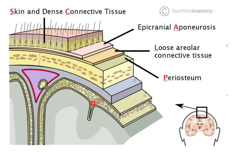 The Scalp Layers Innervation Blood Supply Teachmeanatomy
 Layers Of Skin How Many Diagram Model Anatomy In Order
 Skin Information Layers Of Skin Keeping Skin Healthy And More
 Eps Vector Young And Older Skin Stock Clipart
 Skin Layers Diagram
 Skin 1 The Structure And Functions Of The Skin Nursing Times
 Anatomy Of Skin Layers Vector Image 1815105 Stockunlimited


Comments
Post a Comment