Skin Layers Histology
The organ constitutes almost 8 20 of body mass and has a surface area of approximately 16 to 18 m2 in an adult. Undoubtedly the skin is the largest organ in the human body.
 5 Layers Of The Epidermis 1 Stratum Corneum 2 Stratum
Introduction to skin histology the skin is considered the largest organ of the body and has many different functions.
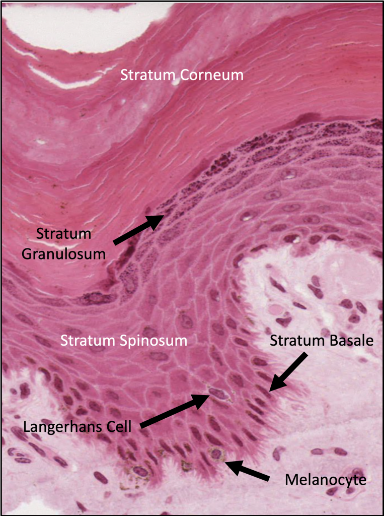
Skin layers histology. This article will describe the anatomy and histology of the skin. Three layers of skin. This epidermis of skin is a keratinized stratified squamous epithelium.
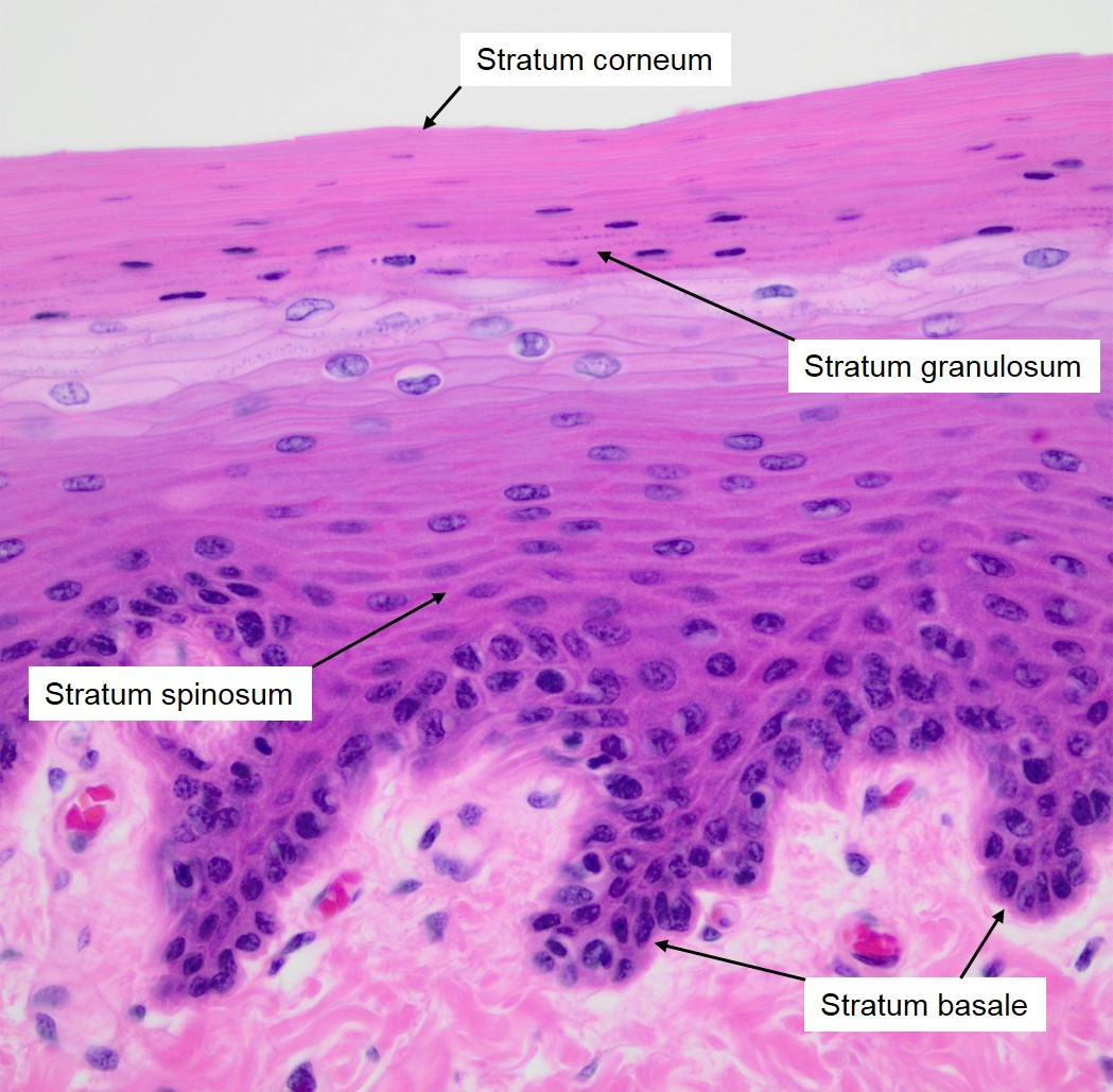
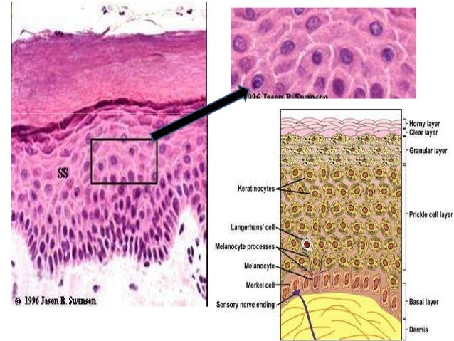
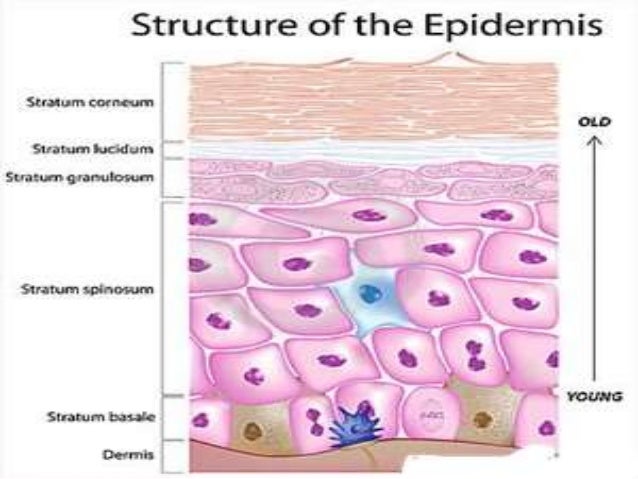
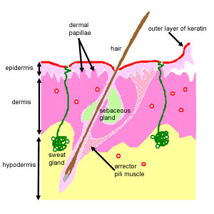
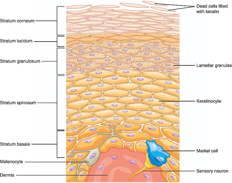
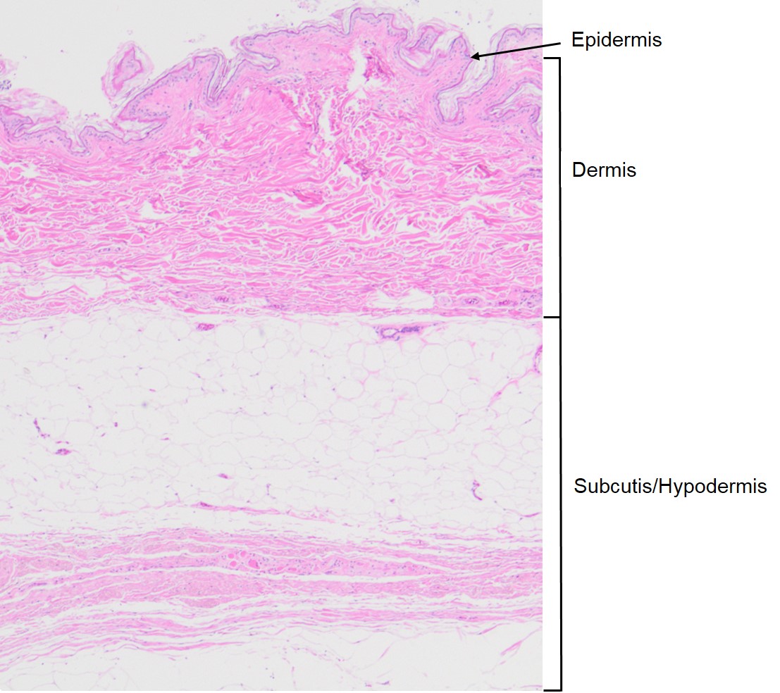
This diagram shows schematically the four different layers found in the epidermis of most skin thin skin. The dermis is attached to an underlying hypodermis also called subcutaneous connective tissue. Layers in the epidermis.
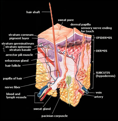
Basic skin histology the skin is divided into two main regions the epidermis and the dermis. The basal layers of this epithelium are folded to form dermal papillae. A thin outer portion that is the keratinised stratified squamous epithelium of skin.
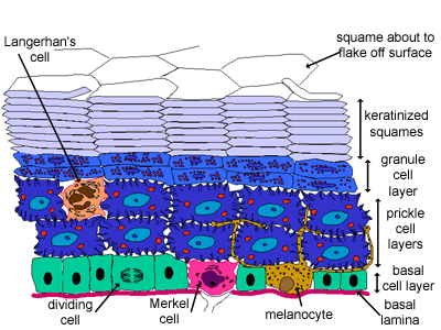
Literally covering you from head to toe. Thin skin contains four types of cellular layers and thick skin contains five. Cells divide in the basal layer and move up through the layers above changing their appearance as they move from one layer to the next.
The epidermis is important for the protective function of skin. Basic skin histology 1.
 Histological Skin Structure Diagram Do It Easy With

 Stratum Lucidum Wikipedia
 Histology Of The Skin

 Pin By Maria Kozlova On Biology Medicine Skin Anatomy
 Structure Of The Epidermis Veterinary Histology
 Basic Skin Histology
 Normal Skin Histology
 Various Layers Of Skin Are Noted In This Histology Slide Of
 Normal Skin Histology Dr Sujeet Kumar

 Histoquarterly Skin Histology Blog
 Is Only The Top Layer Of Skin Cells Alive Socratic
 Skin Histology Flashcards Quizlet

 Histology Finding Of The Normal And Ad Patient S Skin A
 Skin Histology
 112 Best Histology Skin Images Anatomy Physiology
 Integument
 Shotgun Histology Thin Skin
 Skin Medical Student Education Tissupath
 The Histology Guide Skin
![]() Skin Cells Layers And Histological Features Kenhub
Skin Cells Layers And Histological Features Kenhub


 5 1 Layers Of The Skin Anatomy Physiology
The 5 Layers Of Your Skin Dr Leslie Baumann
 Intergumentary System Basics Histology And Effluorescence
 The Histology Guide Skin

 Skin Histology And Microtopography Of Papuan White Snake
 Skin Reading Php Lab

 Histoquarterly Skin Histology Blog
 Basic Skin Histology
 Histological Section Showing Skin Morphology Of A Primitive
 Anatomy Gross Anatomy Physiology Cells Cytology Cell
Histology Of Skin Faculty Of Medicine
 5 1 Layers Of The Skin Anatomy Physiology
 Integument
9 Slide 4 Axillary Skin Aoana02t2 Studocu
Diapositiva 1
 Thick Skin Labeled Histology Thick Skin Mitosis Color
 Characterization Of The Human Ridged And Non Ridged Skin A
 9 Slide 4 Axillary Skin Aoana02t2 Studocu
 Introduction Dermatology Ppt Download
 Human Skin Wikipedia
 Tv2001 2016 Histology Skin And Adnexa Veterinary Science
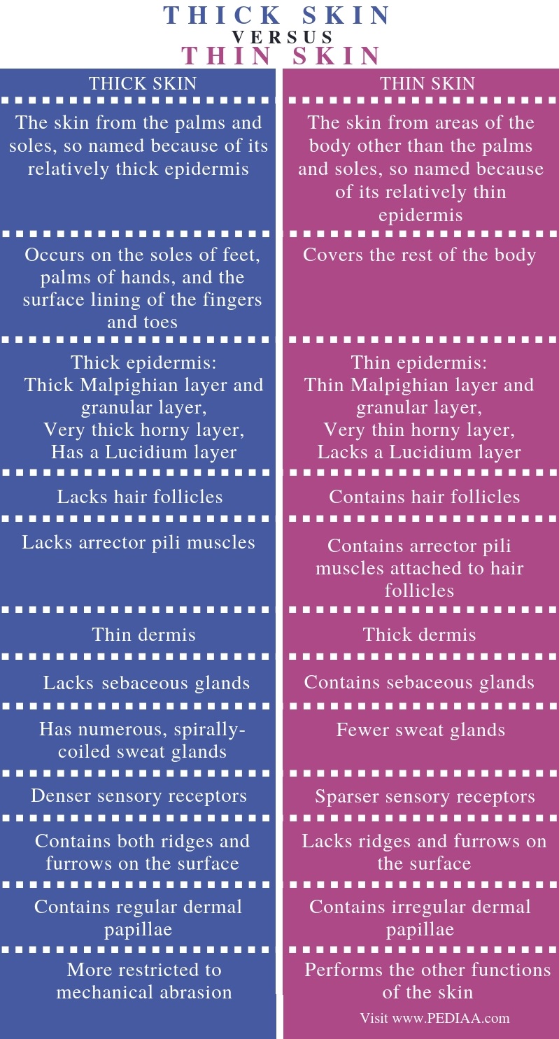 What Is The Difference Between Thick And Thin Skin Pediaa Com
 Normal Histological Structure Of The Epidermis With
 Skin Ultrastructure Epidermis Dermis Teachmeanatomy
 Animal Organs Integument Thin Skin Atlas Of Plant And
 Basic Skin Histology
 Medivisuals Skin Layers Medical Illustration
Skin Thickness As A Potential Marker Of Gestational Age At
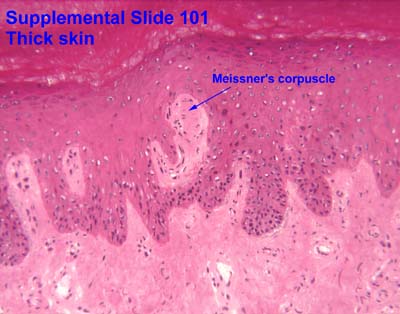
 Figure 2
![]() Skin Cells Layers And Histological Features Kenhub
Skin Cells Layers And Histological Features Kenhub
 Animal Organs Integument Thin Skin Atlas Of Plant And

 Integument Histology Notes Medical Histology Jacobs
 The Histology Guide Skin
Basic Skin Histology
 Normal Skin Histology Embryology Function Blood Supply
 Integumentary System Development Embryology
Diapositiva 1
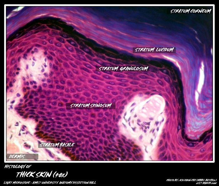 Histology Skin My Aimst University Lifestyle Blog
Plos One Ischemia Reperfusion Injury In A Rat Microvascular
 Skin Histology Skin Layers Epidermis Epidermis Art Skin Layers Watercolor Skin Layers Stratum Corneum Science Art
 Anguilla Anguilla European Eel Skin And Epidermis Vertical Section 200x
 Dictionary Normal Skin The Human Protein Atlas

 Human Skin Atlas
 Hair Follicle 230183665 Image Stock Photo
 Hypodermis Subcutis Subcutaneous Tissue Veterinary
Basic Skin Histology
Aging Skin Histology Physiology And Pathology
 Basic Skin Histology
 Overview Histology Of Skin Structure Showing The 3 Main
 Dictionary Normal Anal Skin The Human Protein Atlas

 Histology World Histology Fact Sheet Skin Appendages
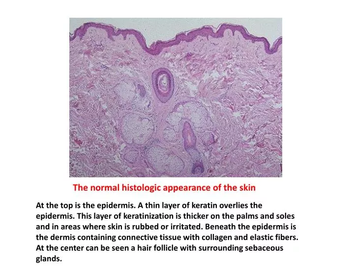 Ppt The Normal Histologic Appearance Of The Skin

 Skin Medical Student Education Tissupath

 Integument
 The Dynamic Natural Skin Care Skin Care Skin Histology
 Histology Skin Part 1 Diagram Quizlet
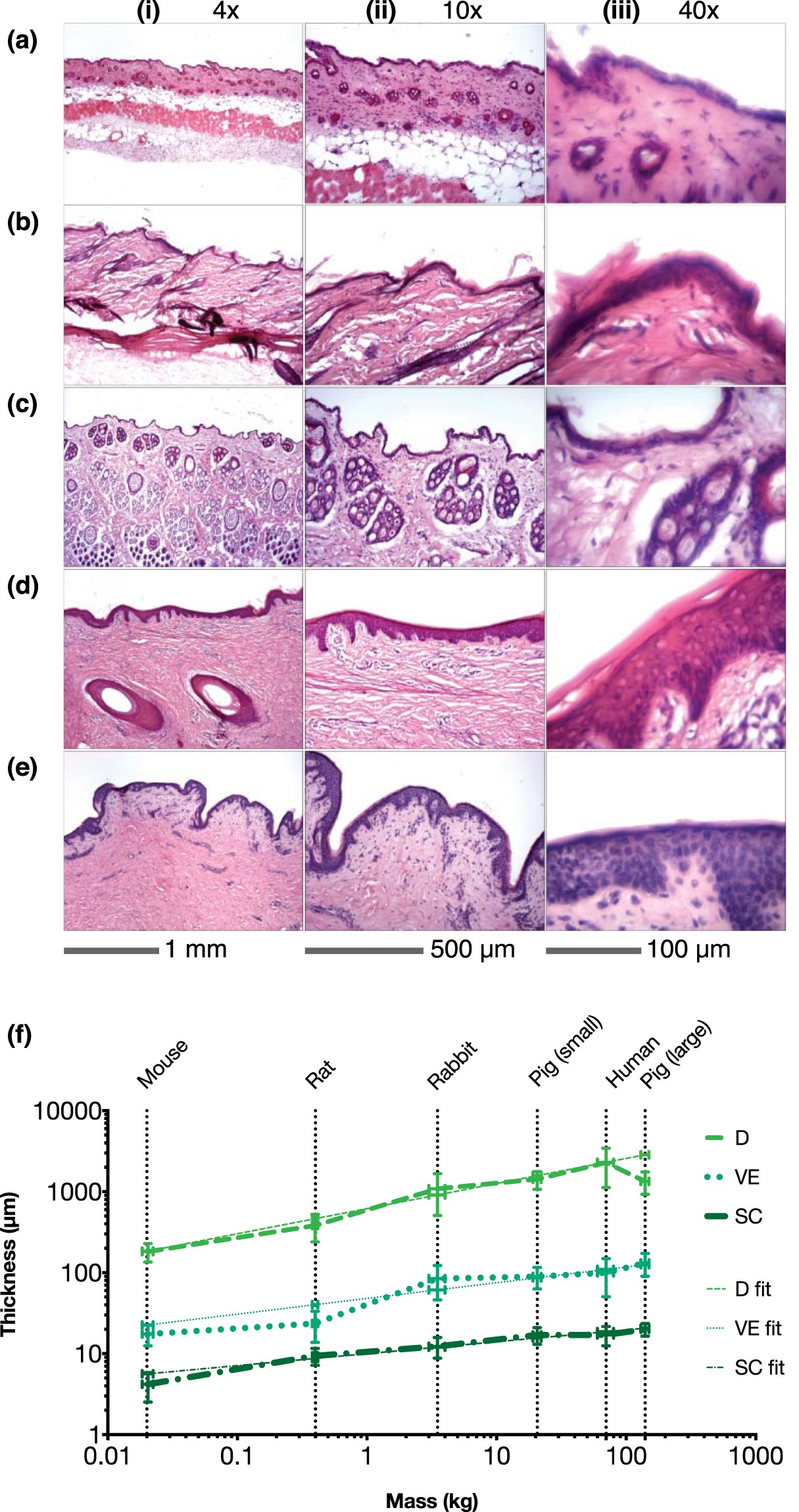 Allometric Scaling Of Skin Thickness Elasticity
 5 1 Layers Of The Skin Anatomy Physiology
Koi Skin
 Histology Of Pig Skin Photography Reprinted From 7 And
 Radio Frequency Assisted Liposuction Rfal Intechopen
 Histology Drawings The Internet Library Of Cortese
 Skin Histology Stock Photos Images Photography Shutterstock

 Mcq On Histology Of Integumentary System Medsynapses
 Bioengineering The Microanatomy Of Human Skin Roger 2019


Comments
Post a Comment